MRI VETS | EXPERTS IN VETERINARY MRI
  • Our Services
  • FAQ
    • For Veterinarians
    • For Animal Owners
  • Contact
  • Online training
  • Animal MRI Centers
  • Community

We read an MRI on a Wallaby today!

10/22/2015

0 Comments

 
Picture
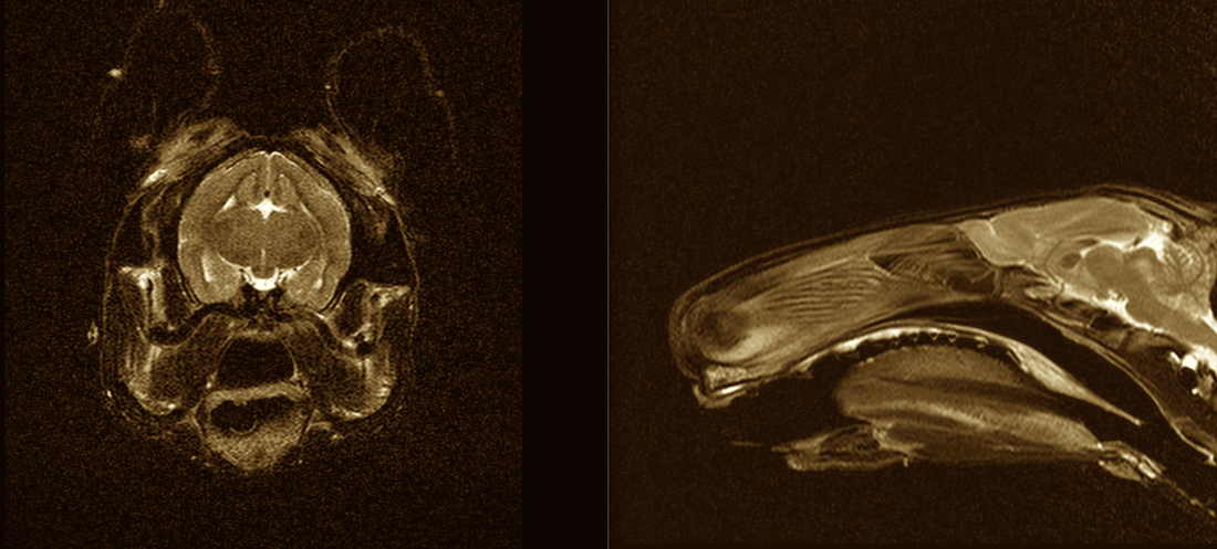
Or maybe that's an Ewok? Cute little guy. And later on in the day we read a CT on an Armadillo!
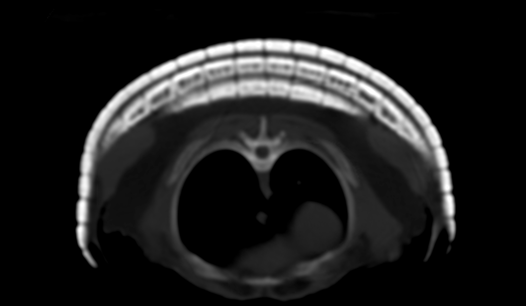
Picture
0 Comments

MRI of Golden Retriever with Seizures 

1/12/2015

0 Comments

 
Golden Retriever Seizures

History

“Penny” is a 7 year-old Golden Retriever. She has been healthy until recently. She has had three seizure episodes in the last month. The seizures appear to be grand mal type with tonic and clonic muscle movements and brief loss of consciousness. Patient appears normal in between the episodes.

MR Examination

Magnetic resonance examination was carried out with a high field, superconducting unit. Sequences included T2 sagittal, T2 transverse, a Fluid Attenuation Inversion Recovery sequence (FLAIR) in the transverse plane, and T1 before contrast in the transverse plane. Gadolinium contrast agent was administered and T1 post-contrast images were obtained in all three planes. Each sequence has approximately 20-25 images and Figs 1-7 illustrate one image per sequence.

Interpretation

All sequences revealed normal anatomy and bilateral symmetry. The mild asymmetric changes within the lateral ventricles are commonly seen as a normal finding in this species. Magnetic resonance imaging has the ability to see macroscopic structural lesions larger than a grain of rice. Therefore, macroscopic structural lesions have been ruled out in this case. The seizures could be due to microscopic or physiologic/metabolic etiologies.

Discussion

Magnetic resonance imaging is an expensive diagnostic test. The value of the test is also high. Dogs that are 7 years of age, when they have their first seizure, could have numerous diseases. The most common etiologies would include infectious conditions, cerebral infarction (strokes), brain tumors, or delayed onset of epilepsy.

An accurate diagnosis is needed for the proper therapeutic options. Early diagnosis can lead to improved treatment outcomes. Placing animals on anti-seizure medication without a proper diagnosis exposes the animal to medications that have significant serious side-effects. In this case, in the absence of a macroscopic finding, anti-seizure medication appears warranted. Proper therapy requires diligence in administering the medication and having blood levels checked for therapeutic levels. The veterinarian and owner can be confident that macroscopic structural lesions, including infarctions, infections, and tumors have been ruled out.

A proper study requires numerous sequences, as illustrated in this case. In addition, the interpretation needs to be done by a skilled professional with experience and the ability to distinguish pathologic change from similar appearing lesions that could be artifactual.

Picture
Picture
Picture
Picture
Picture
Picture
Picture
0 Comments

MRI of Golden Retriever Paw

1/12/2015

0 Comments

 
MRI on Golden Paw

History

“Baxter” was a 2 year-old Golden Retriever. Baxter had a draining tract in his paw that had been surgically explored on three different occasions. Small pieces of wood were found, but there had been no resolution of the fistulous tracts. Dr. Haburjak, a veterinary surgeon, requested a magnetic resonance imaging study of the paw to better plan any further surgical intervention. The previous surgeries would have distorted the normal architecture severely and would make subsequent surgery difficult without an accurate “roadmap.” The magnetic resonance imaging study was carried out with a high field super-conducting magnet. 

Sequences included STIR sequences in all three planes and T1-weighted images before and after contrast. The T1-weighted images were also performed with fat-saturation toreduce the fat signal and allow better visualization of the contrast enhancement.

Interpretation

An abnormality was seen that was within the soft tissues of the paw. The abnormality was between the deep digital flexor tendons and the metacarpal bones of the left paw. There was a hyperintense lesion that was seen on the STIR sequences from a fluid collection. The draining fistulous tracts were readily identified. At the center of the fluid was a hypointense structure that measured approximately 3.5 cm in length by 0.5 cm in width. There was a center to this material, which was compatible to a stick with a soft core or medullary cavity that was soaking up the fluid. 

The lesion was midline between the third and fourth metacarpal regions and was deep to the flexor tendons. Surgery was performed and a stick of the size diagnosed by magnetic resonance was readily removed with the accurate roadmap for the surgeon. The images depict the abnormalities. There is also a post-operative image of the removed wood foreign body. 

The use of magnetic resonance imaging provided the information needed for the proper and corrective surgery. MRI exams are costly, but if one calculates the direct and indirect cost of the previous 3 surgeries there was obviously a value to this examination. When we think of the pain and suffering, the value of a proper procedure often trumps the initial cost.

Images courtesy of Iams Pet Imaging Centers

Picture
Picture
Picture
0 Comments

MRI of Jack Russell with Hind Limb Weakness

1/12/2015

0 Comments

 
Hind Limb Weakness in Jack Russell

History

“Jackson” is a five year old Jack Russell Terrier. Jackson presented with an acute onset of hind limb weakness four days prior to the MR examination. There was a mild improvement initially and then a rapid deterioration. On examination, there was no pain.

Technical Factors

Sequences included T2-weighted sequences in sagittal and transverse planes, STIR sequences in sagittal and dorsal planes, and an MR myelogram.

Interpretation

A readily visualized disc herniation is seen at T12-13. This disc herniation can be seen in figure 1, the T2-weighted sagittal sequence. The fact that there is spinal cord compression is readily seen with the MR myelogram in figure 2. An MR myelogram is a technique to see only the cerebral spinal fluid. No injections are needed. This procedure is totally safe and without the pain and seizures that can accompany conventional myelography.

On figure 3, the herniated disc material can be seen in the ventral and right parasagittal spinal canal. There is loss of the subarachnoid space and spinal cord deformity. The presence of a normal seven lumbar vertebrae with the disc herniation at T12-13 can be confirmed on figure 4. These studies are necessary, as dogs can have 6, 7, or 8 lumbar vertebrae, which would change the surgical approach. Assumption of 7 lumbar vertebrae can be erroneous and lend to surgical problems.

Magnetic resonance imaging for the diagnosis of intervertebral disc herniation has greatly improved clinical outcomes. Contrast myelography, which creates inflammation, adds pain and a degree of complication can now be avoided. In addition, magnetic resonance imaging allows for the clear visualization of the herniated material, facilitating the surgical approach. Contrast myelography had a high error rate as to the side of the herniated disc material. With magnetic resonance imaging, the correct side can be chosen to facilitate complete disc removal and better surgical outcome.

It is our belief that since we have stopped doing conventional contrast myelography, the patients do better following surgery and leave the hospital quicker with a better return to function.

Picture
Picture
Picture
Picture
0 Comments

MRI of German Shepherd with Lameness

1/12/2015

0 Comments

 
Lameness in Shepherd

History

“Max” is a 7 year-old, German Shepherd. Max presents, having had lumbosacral surgery 1 year previously. Max has lameness in the right hind leg at this time. No drawer sign can be elicited but a partial cranial cruciate tear is suspected.

Technical Factors

Sequences include T2-weighted sequences, STIR sequence, and T1-weighted sequences. All planes are represented.

Interpretation

A small amount of joint effusion is seen. For the most part, normal anatomy is visualized. Critical structures, including the cranial and caudal cruciate ligaments, medial and lateral menisci, long digital extensor tendon, straight patellar ligament, and femoral meniscal ligament are visualized and are found to be normal. Figure 1 shows a T1-weighted sagittal image of the stifle with the caudal and cranial cruciate ligament labeled. Figure 2 is a STIR sequence in the same location. The synovial fluid is bright white and the infrapatellar fat body has been suppressed and is now dark. There is excellent contrast between the fat body and the synovial fluid on this sequence. Again, the cranial and caudal cruciate ligaments have been labeled.

The cranial cruciate ligament is never seen as well as the caudal cruciate ligament due to its fan-shaped origin and the volume average with the synovial fluid that envelopes the cranial portion of the ligament. Figure 3 is a T2-weighted image of the stifle medial of midline. The cranial and caudal horns of the meniscus are labeled. Damage to the meniscus is readily detected via magnetic resonance imaging. Figure 4 is a dorsal plane looking at the knee from the front. The lateral and medial collateral ligaments are identified with the arrows. No abnormalities are seen.

The fact that only a mild joint effusion is seen indicates that this animal would not benefit from surgery. Partial tears of the cruciate ligament are readily identified on magnetic resonance imaging as the tear extends into the bone and an abnormality can be seen at the origin and insertion of the ligament. Since that was not detected, this animal merely has a mild degree of osteoarthrosis of the stifle (knee joint).

As our physician colleagues have found, magnetic resonance imaging prior to stifle surgery often indicates that there is not a need for invasive surgery. Procedures to correct stifle instability are both expensive and create significant pain with the potential for complications. Magnetic resonance imaging prevented Max from having unwarranted surgery. While cruciate ligament ruptures and meniscal damage are common in the dog, magnetic resonance imaging can be utilized to confirm that diagnosis prior to surgery and if it is not present, will negate the need for surgery.

Picture
Picture
Picture
Picture
0 Comments

MRI of Dachshund with Paresis

1/12/2015

0 Comments

 
Dachshund with Paresis

History

“Francois” is a 7 year-old Dachshund. Francois presents with a two month history of minimal jumping and over the past 3 days has become very painful and cries when touched. Patient continues to eat and drink, etc. There are no neurologic deficits and no ataxia. Collapse of the L3-4 intervertebral disc is seen. A disc herniation is suspected.

Technical Factors

Sequences include T2-weighted sequences, STIR sequences, and T1-weighted images before and after contrast. T1-weighted images are performed post-contrast with fat-saturation to reduce the bright signal from the fat and allow better visualization of the contrast enhancement.

Interpretation

The abnormality at L3-4 is readily visualized. On figure 1, a sclerotic reaction can be seen involving the caudal endplate of L3 and the cranial endplate of L4. On figure 2, the STIR sequence reveals a bright increased signal intensity at the intervertebral disc space. This is atypical for a dachshund that tends to desiccated intervertebral discs, as seen in the other sites. Also, notice that this animal has an enlarged prostate gland.

Figure 3 is a sagittal image following the administration of gadolinium contrast agent. Contrast enhancement can be seen in the intervertebral disc space. Notice how the fat has turned dark (fat saturation technique) allowing better visualization of the contrast enhancement.

On figure 4, the contrast enhancement of the disc space, as well as in some of the paraspinal musculature, can be readily seen.

This animal has evidence of discospondylitis, an infectious condition of the disc space and intervertebral endplates. This was not detected on a radiographic examination. Only the narrowness of the disc space was detected. This condition can often be confirmed via culture of the blood, CSF, or urine. Most clinicians opt for urine culture. Most common cause of this condition is from a staphylococcus infection from the genitourinary tract. Other conditions, including Brucella sp., have been found. This should always be considered due to the potential for a zoonotic spread to the owner. In this case, Staphylococcus aureus was cultured from the urine.

MR allowed for the definitive diagnosis of discospondylitis in this case. The case went from the typical dachshund with presumed intervertebral disc disease to an animal having an infectious condition. The study prevented this animal from having an unwarranted surgery of the spine and allowed for the condition to be properly treated with antimicrobial therapy. While discospondylitis can cause erosion of the vertebral endplates, that takes time. It takes a 30-50% decrease in bone density in order to be seen radiographically. MR is must more sensitive for the detection of the soft tissue as well as osseous abnormalities.
Just because it’s a dachshund with back pain does not mean it is a disc herniation. Dachshunds, unfortunately, are subjected to all the other diseases known to affect the spine, spinal canal, and spinal cord.


Picture
Picture
Picture
Picture
0 Comments

    Archives

    October 2015
    January 2015

    Categories

    All
    Dachshund
    Foreign Body
    German Shepherd
    Golden Retriever
    Jack Russell
    Lameness
    Limb Weakness
    MRI
    Paresis
    Seizures

    RSS Feed

    Connect with Dr. Gavin

    View my profile on LinkedIn

    Connect with Dr. Sage

    View my profile on LinkedIn
  • Our Services
  • FAQ
    • For Veterinarians
    • For Animal Owners
  • Contact
  • Online training
  • Animal MRI Centers
  • Community