
Case Studies: Imaging Strategies for Localizing Spinal Cord Lesions in Dogs

High-resolution MRI provides critical insight for neurologic cases with subtle or progressive signs.
As a veterinary specialist, you should approach every neurologic case with two guiding questions: (1) What are we seeing, and (2) what might we be missing?
In the following two case studies, advanced imaging clarified spinal cord pathology that was not evident on initial scans. Each highlights how imaging strategy, tailored to the patient, can uncover what the first scan missed.
At SAGE Teleradiology, our radiologists specialize in interpreting high-field MRI and advanced CT studies. With a focus on diagnostic clarity, clinical collaboration, and meaningful next steps, we help you get the full picture when imaging alone isn’t telling the whole story.
[Contact our team] to submit a case and consult with our board-certified specialists today!
In This Article:
Case 1: “Gunner” — Localizing Neurologic Signs with Contrast CT
Case Overview
Initial Workup & Diagnostic Challenges
Advanced Imaging Findings
Clinical Outcome & Impact
Case 2: “Winston” — Progressive Myelopathy Uncovered with MRI
Case Overview
Initial Imaging & Diagnostic Uncertainty
Follow-Up Imaging with Improved Sequences
Clinical Outcome & Impact
Case 1: “Gunner” — Localizing Neurologic Signs with Contrast CT
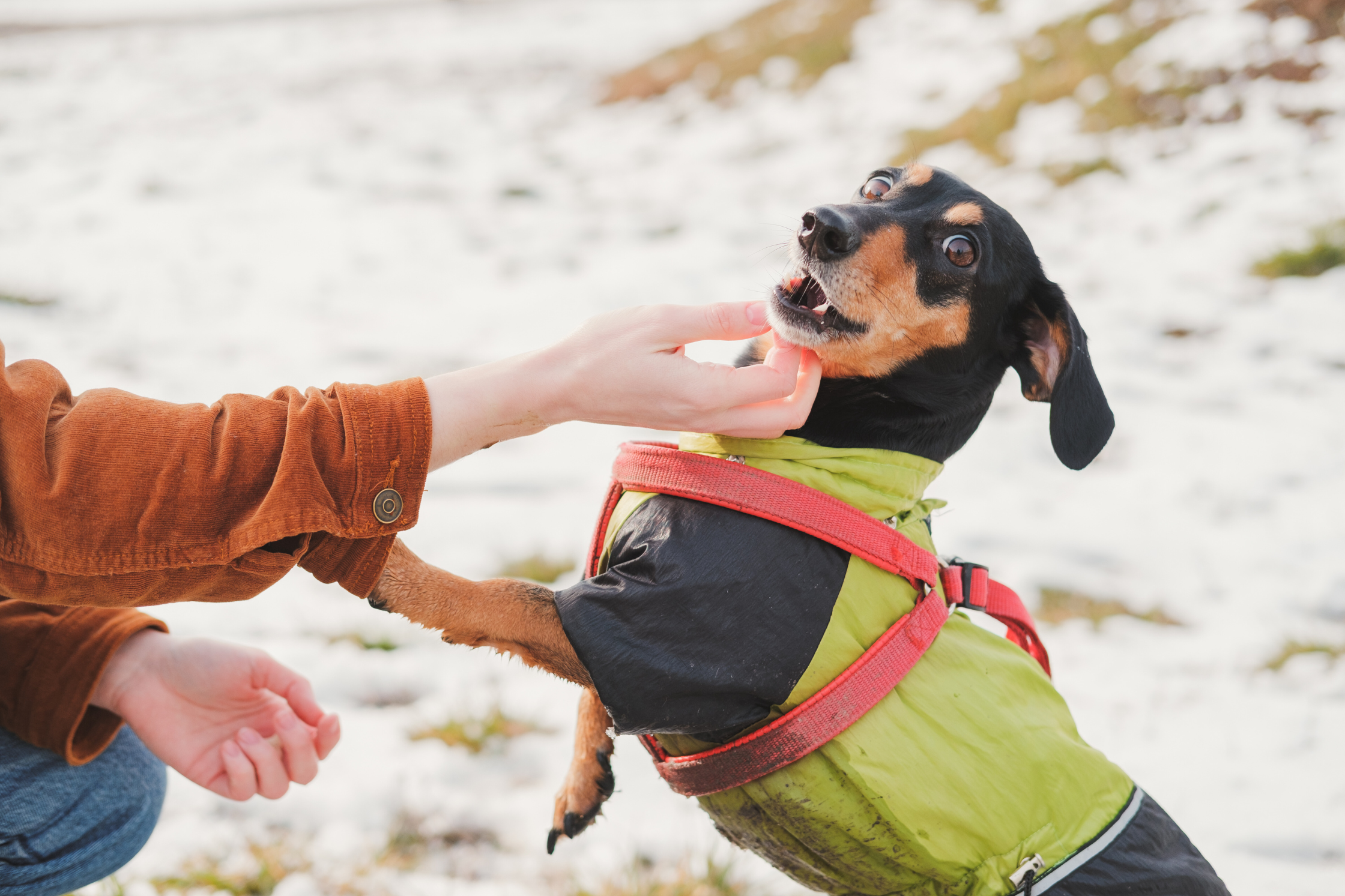
Dachshunds are predisposed to spinal conditions such as intervertebral disc disease and intramedullary lesions.
Case Overview
Patient: Gunner, 8-year-old intact male Dachshund
History: Two-month history of decreased activity and reluctance to move the neck
Exam Findings: No cervical pain; full range of motion
Neurologic Signs: Conscious proprioceptive deficits in both hind limbs; mild to moderate hindlimb ataxia
Suspected Localization: Cervical spinal cord (C1-C5)
Gunner presented with a two-month history of mild but progressive neurologic deficits. Although the owner noted discomfort near the neck, examination revealed no pain and full range of motion. However, his pelvic limb proprioception and coordination suggested a cervical spinal cord lesion.
What made this case notable wasn’t the severity of signs, but the quiet persistence of findings that didn’t yet show up on imaging.
Initial Workup & Diagnostic Challenges

Without contract, the initial CT scan of Gunner shows no visible spinal cord lesion.
A non-contrast CT scan was performed to evaluate for compressive lesions or obvious structural abnormalities. The study was interpreted as normal, with no signs of intervertebral disc disease, vertebral malformation, or mass effect. Given the subtlety of the clinical signs and absence of compressive pathology, localization remained uncertain.
Orthopedic causes were briefly considered, but the neurologic pattern and progressive course kept the focus on the spinal cord. With clinical suspicion still high and no definitive lesion identified, repeat imaging with IV contrast was pursued. The goal was to assess for intra-axial pathology, such as inflammation or neoplasia, that may not be visible without enhancement.
Advanced Imaging Findings

Contrast-enhanced CT reveals a clear spinal cord lesion in Gunner’s cervical spine.
The contrast-enhanced CT revealed a focal area of enhancement within the cervical spinal cord parenchyma—an intramedullary lesion consistent with neoplastic or inflammatory disease.
It was well-demarcated, non-compressive, and not associated with structural distortion, explaining its absence on the non-contrast study.
This single adjustment in imaging strategy provided three key clinical benefits:
It confirmed the suspected localization.
It clarified the nature of the lesion.
It gave the clinical team actionable direction.
✎ Radiologist’s Note:
Persistent neurologic signs with non-revealing imaging shouldn’t be dismissed—they often reflect pathology that standard techniques can’t resolve. In this case, contrast wasn’t a backup plan. It was the step that made the diagnosis possible.
Clinical Outcome & Impact
With the lesion identified, the referring clinician was able to rule out orthopedic causes and confidently localize the issue to the cervical spinal cord. This led to a targeted discussion with the client about next steps, including referral for advanced neurologic evaluation and the potential for medical management of a presumptive inflammatory or neoplastic process.
Early identification allowed the clinical team to start corticosteroid therapy while awaiting referral, providing symptom relief and giving the client a clearer understanding of prognosis and options.
Case 2: “Winston” — Progressive Myelopathy Uncovered with MRI

Airedale Terriers are active, large-breed dogs that can develop age-related spinal cord disease.
Case Overview
Patient: Winston, 11-year-old neutered male Airedale Terrier
Chief Complaint: Progressive hindlimb weakness evolving to paraparesis
Exam Findings: Declining pelvic limb strength with coordination deficits
Clinical Course: Ongoing neurologic decline despite stable general health
Initial Concern: Suspected thoracolumbar spinal cord lesion
Winston presented with a slowly progressive decline in pelvic limb function. While his general health remained stable, the neurologic deficits had become increasingly apparent. The absence of spinal pain added complexity to the clinical picture, but the pattern suggested a central spinal cord process affecting the thoracolumbar region.
Given his age and the chronic, non-traumatic onset, the clinical team considered a range of differentials—including inflammatory, neoplastic, or vascular disease—and advanced imaging was pursued to clarify the diagnosis.
Initial Imaging & Diagnostic Uncertainty

Initial MRI of Winston’s spine with limited resolution; no clear lesion was identified.
A low-field MRI was performed as the initial imaging modality. While it revealed vague intramedullary changes on T2-weighted sequences, no discrete lesion or compressive abnormality was identified. With localization unclear and no definitive pathology seen, diagnostic uncertainty remained.
In cases like Winston’s, where neurologic signs evolve slowly and imaging results are inconclusive, the absence of a definitive finding doesn’t rule out disease. It simply calls for further optimization: repeat imaging, improved sequence selection, or higher-field MRI.
Follow-Up Imaging with Improved Sequences

Follow-up MRI reveals a well-defined spinal cord lesion consistent with progressive disease.
A high-field MRI was performed in the following days, utilizing optimized sequences and improved resolution. This scan revealed a well-demarcated intramedullary T2 hyperintensity, consistent with spinal cord disease, now clearly visible on both sagittal and axial images.
While not dramatic in size, the lesion correlated directly with the clinical picture. The enhanced imaging clarified localization and gave the veterinary team a solid basis for treatment planning and client communication.
✎ Radiologist’s Note
In evolving spinal cord disease, the first scan may not tell the full story. Winston’s case highlights the diagnostic value of persistence—not through repetition for its own sake, but through intentional refinement of technique and timing.
Clinical Outcome & Impact
The updated MRI provided clarity in lesion localization and allowed the referring veterinarian to have a more informed discussion about prognosis and treatment options. Whether pursuing advanced referral or initiating supportive care, the team could now move forward with confidence.
Winston’s case is a reminder that repeat imaging isn’t a fallback. It’s a forward step when clinicals continue to evolve. A second look can make the difference between diagnostic uncertainty and actionable insight.
Key Takeaways for Clinicians
Initial imaging, especially non-contrast or low-field studies, may not reveal subtle spinal cord lesions.
When neurologic signs persist or progress, contrast-enhanced CT or repeat MRI with optimized technique can improve diagnostic yield.
Subtle deficits without overt pain should raise suspicion for intramedullary disease and prompt advanced imaging.
Strategic imaging decisions–modality, timing, and sequencing—can turn diagnostic uncertainty into clinical clarity.
In spinal imaging, quality is cumulative: what you see depends not just on the scan, but how and when it’s performed.
Whether you're localizing a vague myelopathy or tracking progressive deficits, these examples reinforce how timing, technique, and image quality drive diagnostic confidence.
✎ Radiologist’s Note
Imaging doesn’t happen in a vacuum and neither should interpretation. A scan is just one piece of the puzzle. When you combine high-quality imaging with clinical insight, physical exam findings, and supporting diagnostics, you get more than a picture. You get a plan—and that’s what drives better outcomes.
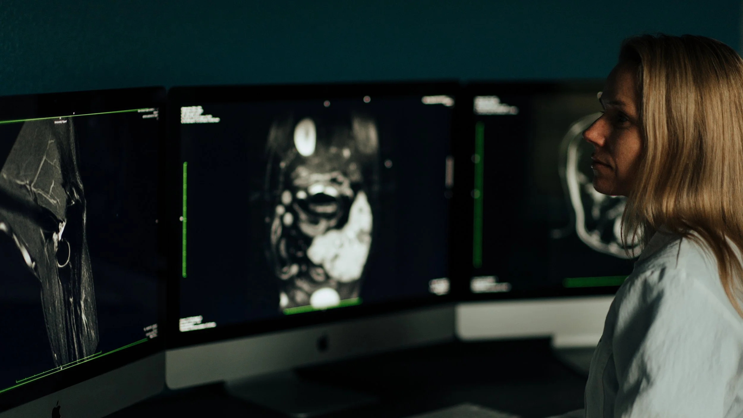
Lead radiologist, Dr. Jaime Sage, reviews an MRI scan at Sage Veterinary Imaging’s Texas location.
Have a Challenging Neuro Case? Let’s Take a Second Look.
Our radiologists specialize in interpreting high-field MRI and advanced CT studies with a focus on diagnostic clarity, clinical collaboration, and meaningful next steps. When imaging alone isn’t telling the whole story, we help you get the full picture.
[Contact our team] to submit a case and consult with our board-certified specialists today!